|
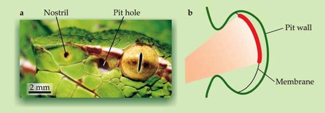
Two families of snakes, pit vipers and boids, possess an infrared (IR) sensitive sensory system that is used to construct a two-dimensional representation of the heat distribution in their environment. In the case of the pit viper, the sensory organ consists of two cavities (pit holes) lying closely to the eyes at the sides of the head (figure 1a). Inside this cavity a freely suspended membrane (1b) is contained. The membrane is very thin (15 micrometer) and contains heat-sensitive cells that can detect temperature differences down to a few mK. Still, the opening of the pit hole must be large, to be able to quickly detect moving prey. Therefore, the optical quality of the organ as "measurement device" is very poor. Nontheless, the snake is able to precisely strike prey using the heavily blurred heat image on the pit membrane (figure 3a). This paradox has been resolved.
|
Figure 1: (a) This profile of a pit viper shows the opening of the left pit hole. The cavity lies between the nostril and the eye. The schematic picture (b) shows the proportions of the relevant sizes. The opening of the pit organ is about as large as the depth of the organ. Because of this, a heat point in space will be projected onto a smeared out patch on the membrane. An image of the environment is not recognizable anymore (Figure 3a). [All figures from Physics Today.]
|
|
We investigated how the snake is able to form an accurate neuronal image of the environment. To this end we developed a model that "replaces" the non-existing lens by a neuronal processing algorithm. A virtual lens in the snake brain.
|
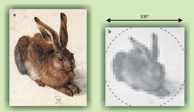
Figure 2: (a) Albrecht Dürer's Hare from 1502. (b) A pixelized (32x32) version of the hare was used as an exemplar heat distribution. The dotted line shows the view field of the pit organ with a width of ca. 100 degrees.
|
|
As "prey animal", the hare by Dürer (figure 2a) was used. Our model reconstructs every single pixel of the prey animal from the measured heat distribution on the membrane (figure 3a). The mathematical operations that are needed to accomplish this can be easily performed in a neuronal feed-forward network. A closer investigation of the algorithm showed that the underlying mechanism of the reconstruction consists of a set of edge detectors. Similar edge detector systems are found for instance in the visual system, where they contribute to contrast enhancement and sharpening. Biologists have found a neuronal map of IR input in the snake optic Tectum, which confirms our idea that a neuronal input reconstruction is formed in the snake's brain.
|
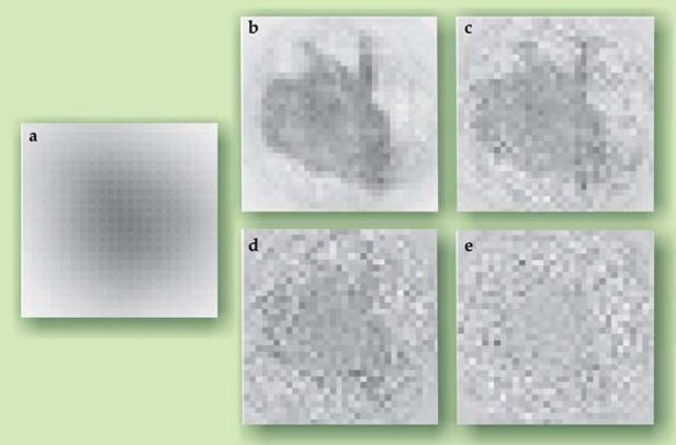
Figure 3: For the challenge to reconstruct the hare from figure 2b, only the measurements from the pit membrane (a) are used. The panels (b-e) show the reconstruction using our model for different values of the detection noise (0.25%, 1%, 2% and 5%).
|
|

last modified 2007-11-05
by
webmaster@Franosch.org
|